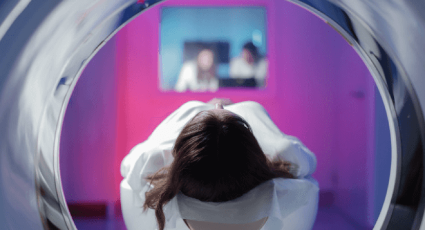
An extremity MRI is a type of MRI used to examine the extremities’ bones, muscles, tendons, ligaments, and joints.
An extremity MRI in Sparta, NJ, is done for patients who have pain in their arms or legs and would like to know if there are any problems with their joints. The patient will be scanned from the wrist up to the elbow or knee.
More about extremity MRI
MRI is a technology that has been used to diagnose, monitor, and treat various diseases. You can also use it in other fields such as sports medicine, physical therapy, and neurology. It is a non-invasive procedure that uses strong magnetic fields and radio waves to create body images. It is often used to produce detailed images of soft tissues such as organs, muscles, tendons, or ligaments.
The extremity MRI is a magnetic imaging technique that uses radio waves and the strength of the Earth’s magnetic field to produce detailed images of soft tissue and organs in a short time.
The procedure produces high-quality images with minimal radiation exposure and has many applications in medical fields, including musculoskeletal imaging, cardiology, neurology, neurosurgery, and orthopedics.
Applications of extremity MRI:
Extremity MRI is a type of MRI used to assess the extremities. It is an essential part of a medical exam and can detect injury or disease.
Extremity MRI can diagnose and treat musculoskeletal, neurologic, and vascular conditions. The most common applications are for the following:
- Musculoskeletal conditions: bone fractures, bone tumors, arthritis, joint instability, dislocations/luxation
- Neurologic conditions: stroke/TIA (transient ischemic attack), brain tumors, epilepsy
- Vascular diseases: aneurysms/arteriovenous malformations
- Soft tissue disorders such as tendonitis and carpal tunnel syndrome
- Fractures and dislocations
How long does the procedure of extremity MRI take?
An extremity MRI is a type of MRI used to examine the body’s extremities. It is often used in cases where there is pain, swelling, or deformity in the extremities.
The procedure can take up to 20 minutes. The patient will be asked to lie on their side with their arm below their head and feet. They will be asked not to move during the procedure and may feel some pressure when moving into position for imaging.


